


 4")

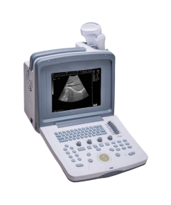
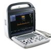
DCU10 Color Doppler Ultrasound System
R91,072.40 Inc VAT
The DCU10 color Doppler ultrasound delivers clear diagnostic imaging with real-time color flow visualisation. A compact, reliable system suited for general practice, specialist clinics, and outpatient settings.
Standard configuration:
| Main unit:1 pc |
| Double probe sockets, automatically identify probe; |
| 3.5MHz multi-frequency abdomen convex probe:1pc |
| 7.5MHz multi-frequency high linear probe:1pc |
| CD: 1pc |
| Reticle: 1pc |
DCU10 Color Doppler Ultrasound System
Why Choose the DCU10 for Your Practice?
USE
For the examine of abdomen, heart, gynecology, obstetrics, urology, small organs, pediatrics, blood vessels, etc.
The DCU10 Color Doppler Ultrasound System offers clinicians a reliable, accessible imaging platform for a broad range of diagnostic applications. With color Doppler capability, it provides real-time visualisation of blood flow patterns — useful in vascular assessment, cardiac screening, and obstetric monitoring.
The system supports multiple probe types, giving it versatility across imaging disciplines without the need for separate equipment. Its processing engine delivers clear, detailed greyscale images enhanced by color Doppler overlays, helping clinicians identify abnormalities quickly and confidently.
Designed for busy clinical environments, the DCU10 combines practical ergonomics with professional-level imaging. The interface is straightforward, reducing scan times and improving workflow efficiency. Whether used in a hospital department or outpatient clinic, it delivers consistent, repeatable results.
Compare with the DCU12 scanner for advanced imaging requirements. For ultrasound training and clinical guidance, explore resources at AIUM.org.
2. Monitor
High resolution 15 “LCD display.
3. Image display mode
B,2B,B+M,4B,M,B+Color,B+PDI,B+PW,B+Color+PW,B+PDI+PW,★B+C/PDI Double real time
4. Probe elements: 128
5.1 Probe specification
| Probe specification | 3.5MHz multi-frequency convex probe | 7.5MHz multi-frequency linear probe | 6.5MHz multi-frequency transvaginal probe |
| Scanning angle | 60° | / | 135° |
| elements | 80 | 80 | 80 |
| frequency scope | 2.0MHz-5.0MHz | 6.0MHz-9.0MHz | 4.5MHz-9.0MHz |
| 4 multi-frequency | 2.0/2.5/3.5/5.0 | 6.0/6.5/7.5/9.0 | 4.5/5.5/6.5/7.5 |
| Max scanning depth | ≥190mm | ≥60mm | ≥60mm |
| Blind zone | ≤5mm | ≤3mm | ≤4mm |
5.2 Operating mode
B,B/B,4B mode
M,B/M mode
CFM
PDI
PW
THI
6. Measurement / Calculation
6.1 General measurement
B Mode measurement: distance, area (distance measurement method, ellipse distance method, trace method), circumstance (distance measurement method, ellipse distance method, trace method), volume (two – axial method), angle , Histogram , sectional view, stenosis rate( length stenosis rate, area stenosis rate ) and depth.
M mode measurement: depth, slope, heart rate, circle
D mode measurement: velocity, heart rate, flow velocity, time acceleration, Pulse/ Resistance Index, Max differential pressure, PG mean,ratio, stroke volume, flow rate.
6.2 Gynecology measurement and analysis: uterus, endometrium, ovary, cervix, follicle.
6.3 Obstetric measurement and analysis
BPD),CRL,GS,FL,HL,TAD,TTD,APTD,HC,AC,EDC,FT,LV,THD,TCD,OFD,GA,EFW,LMP,AFI,and calculate GA, EFW, EDD and growth curve, different formula of many counties.
6.4 Urology measurement and analysis: Prostate volume, Residual Urine Volume
6.5 Cardiology measurement and analysis
6.6 Measurement report:
Obstetric measurement report,
gynecology measurement report,
cardiology measurement report,
urology measurement report and other measurement reports
Automatically store measurement results and generate report
Standard configuration:
| Main unit:1 pc |
| Double probe sockets, automatically identify probe; |
| 3.5MHz multi-frequency abdomen convex probe:1pc |
| 7.5MHz multi-frequency high linear probe:1pc |
| CD: 1pc |
| Reticle: 1pc |
Optional configuration:
| Video Printer(sony 897-MD) |
| 6.5MHz multi-frequency trans-vaginal prob |
Be the first to review “DCU10 Color Doppler Ultrasound System”
Related products


Reviews
There are no reviews yet.